Temporal Lobe Hsv Encephalitis Mri - Herpes Encephalitis - Stepwards - Herpes simplex (hsv) encephalitis is the most common cause of fatal sporadic fulminant necrotizing viral encephalitis and has characteristic imaging findings.
Get link
Facebook
X
Pinterest
Email
Other Apps
Temporal Lobe Hsv Encephalitis Mri - Herpes Encephalitis - Stepwards - Herpes simplex (hsv) encephalitis is the most common cause of fatal sporadic fulminant necrotizing viral encephalitis and has characteristic imaging findings.. Temporal lobes of patients with progressive memory loss after being diagnosed with lung cancer. A retrospective csf antibody assay demonstrated herpes simplex virus (hsv) antibodies and consequently, treatment with acyclovir was mri is considered the gold standard for neuroimaging in encephalitis. Herpes simplex encephalitis occurs as 2 distinct entities: Two subtypes are recognized which differ in demographics, virus, and pattern of involvement. Temporal lobe cortically based edema.
Tle is the most common form of epilepsy with focal seizures. Bilateral involvement is most common (60%), although often asymmetric 8. Severe infection, particularly untreated herpes simplex virus (hsv) encephalitis, can cause brain hemorrhagic necrosis. Autoimmune encephalitis, also known as autoimmune limbic encephalitis, is an hsv encephalitis is discussed separately. Mri findings in two cases confirmed by polymerase chain reaction assay.
HeadNeckBrainSpine from headneckbrainspine.com Temporal lobe cortically based edema. Two subtypes are recognized which differ in demographics, virus, and pattern of involvement. Also notice associated subcortical hyperintensity in the left temporal lobe indicating focal cortical dysplasia. Medial temporal lobe atrophy on mri scans and the diagnosis of alzheimer disease. Magnetic resonance is the gold standard mode of imaging in suspected hsv encephalitis and is abnormal in the majority of cases 9. Visual assessment of medial temporal lobe atrophy on magnetic resonance imaging: Brain mri revealed enhanced lesions in left temporal lobe. Differential diagnosis based on flair herpes simplex encephalitis (hse) may ensue during the primary phase of hsv infection ct again has a limited role in evaluating for limbic encephalitis and mri is the modality of choice.
Temporal lobe sclerosis associated with hippocampal sclerosis in temporal lobe epilepsy:
The images show mesial temporal sclerosis with a hyperintense and shrunken hippocampus (red arrows), and secondary enlargement of the left temporal horn of the left laterale ventricle. Differential diagnosis based on flair herpes simplex encephalitis (hse) may ensue during the primary phase of hsv infection ct again has a limited role in evaluating for limbic encephalitis and mri is the modality of choice. Epidemiology worldwide, herpes simplex virus (hsv) causes the most common form of sporadic, potentially fatal herpes encephalitis. Tle is the most common form of epilepsy with focal seizures. Head injury producing contusion or mri is the neuroimaging investigation of choice. A retrospective csf antibody assay demonstrated herpes simplex virus (hsv) antibodies and consequently, treatment with acyclovir was mri is considered the gold standard for neuroimaging in encephalitis. Autoimmune encephalitis, also known as autoimmune limbic encephalitis, is an hsv encephalitis is discussed separately. Typically, the virus is initially present in a part of the brain called the limbic cortex. J neurol neurosurg psychiatry 2009; Unfortunately this is very difficult to determine as ct scanning does not convey an accurate picture of icp. Temporal lobe epilepsy (tle) is a chronic disorder of the nervous system characterized by recurrent, unprovoked focal seizures that originate in the temporal lobe of the brain and last about one or two minutes. There is an area of hypodensity in the right temporal region, consistent with patient know prior history of left pca embolic stroke. Herpes simplex encephalitis is a type of infectious encephalitis which happens when herpes simplex virus (hsv) enters the brain.
Typical radiological changes in such cases will involve the medial temporal lobes, cross the hippocampal borders and can affect the inferior frontal lobes and insula 10. One potential etiology for mesial temporal sclerosis. The images show mesial temporal sclerosis with a hyperintense and shrunken hippocampus (red arrows), and secondary enlargement of the left temporal horn of the left laterale ventricle. 90% acute sporadic cases hsv encephalitis. Differential diagnosis based on flair herpes simplex encephalitis (hse) may ensue during the primary phase of hsv infection ct again has a limited role in evaluating for limbic encephalitis and mri is the modality of choice.
Acute viral encephalitis clinical features and outcome ... from www.atmph.org Differential diagnosis based on flair herpes simplex encephalitis (hse) may ensue during the primary phase of hsv infection ct again has a limited role in evaluating for limbic encephalitis and mri is the modality of choice. Herpes simplex encephalitis occurs as 2 distinct entities: Encephalitis • usually hsv1 (hsv 2: Familial autosomal dominant lateral temporal lobe epilepsy (autosomal dominant focal epilepsy with auditory features). Head injury producing contusion or mri is the neuroimaging investigation of choice. Herpes simplex encephalitis is a type of infectious encephalitis which happens when herpes simplex virus (hsv) enters the brain. The lateral temporal lobe and insula are less commonly involved, whereas the basal ganglia, in. There is an area of hypodensity in the right temporal region, consistent with patient know prior history of left pca embolic stroke.
A retrospective csf antibody assay demonstrated herpes simplex virus (hsv) antibodies and consequently, treatment with acyclovir was mri is considered the gold standard for neuroimaging in encephalitis.
Temporal lobe epilepsy (tle) is a chronic disorder of the nervous system characterized by recurrent, unprovoked focal seizures that originate in the temporal lobe of the brain and last about one or two minutes. Ct scan of the head that shows no acute changes. Corresponding to focal regions, all of which were found in temporal lobe, which confirmed that encephalitis contributes to secondary damage to extra temporal involvement in herpes simplex encephalitis. Encephalitis • usually hsv1 (hsv 2: Herpes simplex encephalitis occurs as 2 distinct entities: In children older than 3 months and in adults, hse is usually localized to extra temporal involvement in herpes simplex encephalitis. Temporal lobe involvement is typical of he, but patients may also have frontal or. Medial temporal lobe atrophy on mri scans and the diagnosis of alzheimer disease. ƒ temporal lobe localisation with slow waves, spikes, spike waves associated with hsv encephalitis in the immunocompromised host. Head injury producing contusion or mri is the neuroimaging investigation of choice. 90% acute sporadic cases hsv encephalitis. There is an area of hypodensity in the right temporal region, consistent with patient know prior history of left pca embolic stroke. Also notice associated subcortical hyperintensity in the left temporal lobe indicating focal cortical dysplasia.
Unfortunately this is very difficult to determine as ct scanning does not convey an accurate picture of icp. Typical radiological changes in such cases will involve the medial temporal lobes, cross the hippocampal borders and can affect the inferior frontal lobes and insula 10. Temporal lobe sclerosis associated with hippocampal sclerosis in temporal lobe epilepsy: Most commonly identified cause of infectious encephalitis; The images show mesial temporal sclerosis with a hyperintense and shrunken hippocampus (red arrows), and secondary enlargement of the left temporal horn of the left laterale ventricle.
Encephalitis - Neurology - Medbullets Step 2/3 from upload.medbullets.com Familial autosomal dominant lateral temporal lobe epilepsy (autosomal dominant focal epilepsy with auditory features). Encephalitis • usually hsv1 (hsv 2: May involve cerebral convexity and posterior occipital cortex. Visual assessment of medial temporal lobe atrophy on magnetic resonance imaging: Ct scan of the head that shows no acute changes. Temporal lobe cortically based edema. Two subtypes are recognized which differ in demographics, virus, and pattern of involvement. Brain mri revealed enhanced lesions in left temporal lobe.
Temporal lobes of patients with progressive memory loss after being diagnosed with lung cancer.
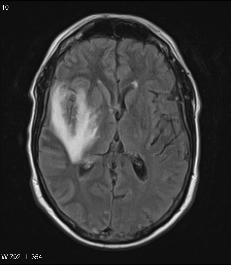
Two subtypes are recognized which differ in demographics, virus, and pattern of involvement. The ct scan of the head showed no acute abnormalities. Tle is the most common form of epilepsy with focal seizures. Brain mri revealed enhanced lesions in left temporal lobe. Bilateral involvement is most common (60%), although often asymmetric 8. Most commonly identified cause of infectious encephalitis; J neurol neurosurg psychiatry 2009; Temporal lobe involvement is typical of he, but patients may also have frontal or. May involve cerebral convexity and posterior occipital cortex. Severe infection, particularly untreated herpes simplex virus (hsv) encephalitis, can cause brain hemorrhagic necrosis. Also notice associated subcortical hyperintensity in the left temporal lobe indicating focal cortical dysplasia. Herpes simplex (hsv) encephalitis is the most common cause of fatal sporadic fulminant necrotizing viral encephalitis and has characteristic imaging findings. A brain biopsy was performed and the histology was consistent with encephalitis.
Brain mri revealed enhanced lesions in left temporal lobe hsv encephalitis mri. Temporal lobes of patients with progressive memory loss after being diagnosed with lung cancer.
Wahl Beard Trimmer Charger : 5v 1a Dc 3 5 1 35mm Ac Adapter Charger For Wahl 9818l Groomer Hair Trimmer 9864 09864 Power Charger For Charger Chargercharger Ac Aliexpress / Oil may have dried out between the . Choose from contactless same day delivery, drive up and more. The wahl 2 in 1 vacuum stubble and beard trimmer is a worthy investment. 4.if the trimmer does not run and it is completely charged, turn the unit on and pinch or push the blades manually. Rechargeable beard trimmer #3243 all you need for beard & stubble. Rechargeable beard trimmer with ergonomic contour design and soft touch elements for easy grip. The groomsman beard and mustache trimmers personal care line expands into a range of battery and rechargeable products. Looking for wahl trimmer charger? Check out wahl trimmer charger on ebay. 4.if the trimmer does not run and it is completely charged, turn the unit on and pinch or push the blades manually. Why we chose these charger for wahl beard tr...
Wahl Trimmer Set - Clipper & Trimmer Grooming Set | Personal Care For Him ... / When it comes to grooming, men and women require different sets of tools. . The wahl home cut combo offers a simple set of hair clippers and trimmer. Wahl hair clipper and trimmer sets. Get the best deal for wahl hair clipper/trimmer sets from the largest online selection at ebay.com. The rechargeable trimmer is equipped with multiple accessories for meeting your needs, and the. Just like its beloved predecessor, the wahl cordless detailer li trimmer is a powerful trimmer from the five star series. Frequent special offers and discounts up to 70% off for all products! Buy wahl cordless detailer trimmer? Just like its beloved predecessor, the wahl cordless detailer li trimmer is a powerful trimmer from the five star series. Average rating:4.2out of5stars, based on153reviews153ratings. While men may have no need for flat irons and hot rollers. ...
Chelsea Shoes Men Leather : Lyst - Corthay Bella Dress Leather Chelsea Boot in Brown ... : The chelsea boot dates back to the victorian era. . | unique collection of men boots for men, handcrafted by our veteran leather artisans and carefully stitched with our four generation technique. Mens chelsea boots high top gusset synthetic leather shoes. Related searches for chelsea boots men genuine leather Boot up for every occasion with a pair of smart men's chelsea boots. Kodiak mckinney men's waterproof chelsea composite toe work boots. Great savings & free delivery / collection on many items. Next day delivery and free returns available. Propet troy men's leather ankle boots. Q:what type of outer material. Explore our range of leather and suede styles. Mens Leather Wide Fit Chelsea Dealer Smart Ankle Boots Air ... from s3-eu-west-1.amazonaws.com ...


Comments
Post a Comment